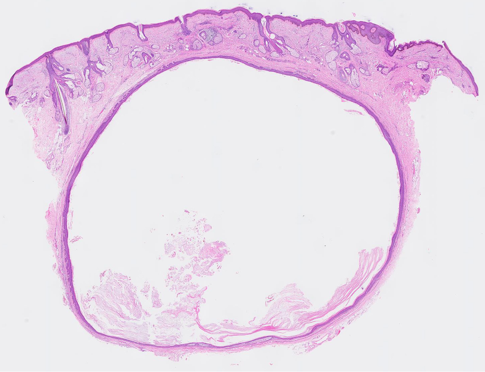
Central Laminated Keratotic Debris

Disease Specific Diagnostics And Medical Management Neupsy Key

Keratosis Obturans Radiology Reference Article Radiopaedia Org

If You Have A Mole At One Of These 7 Places On Your Body This Is What It Means You Will Be Surprised Mole Meaning Natural Medicine Good Interpersonal Skills

Use Once A Week Doubles Hair Growth Combine 1 Tsp Honey 2 Tsp Olive Oil And 1 2 Tsp Coconut Oil W One Half Mashed Avocado Massage Into Dry Hair Wait 10

10 Best Dermatology Images In 2020 Dermatology Wound Care Nursing Medical Knowledge
Nodular And Diffuse Dermatitis Chapter 8 Pearls And Pitfalls In Inflammatory Dermatopathology

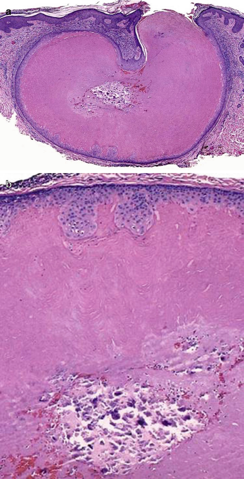
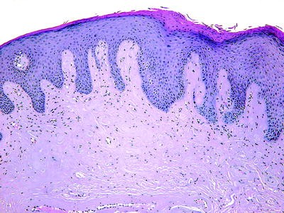
On histologic examination the tumors are composed of an endophytic exophytic cup shaped squamous epidermal proliferation containing a crater like center filled with laminated keratotic material fig.
Central laminated keratotic debris. Presents with pruritic hyperkeratotic and ulcerated nodules and papules with a central keratotic plug mostly located on extensor surface of upper and lower limbs and on the trunk. Left side donate all pages. The absence of hair structure and the presence of inflammatory debris within the invaginations were highly suggestive of kyrle s disease. Actinic keratosis a sharply outlined wartlike or keratotic growth which may develop into a cutaneous horn and may become malignant.
Keratosis ker ah to sis any horny growth such as a wart or callosity. Electron microscopic studies of a follicular lesion showed extracellular viral particles suggestive of a polyomavirus within the central follicular keratotic debris. Keratin is a fibrous protein found in nails hair and the outer layer of skin. Dna polymerase chain reaction pcr and gene sequencing studies performed on the tissue of the microscopic slide and paraffin block for the recently identified ts associated.
Subsequent re epithelization from the adjacent epidermis covers this entire process from the base. Two pyogenic granulomas arising from the wall of an epidermoid cyst on the midback of a 62 year old white man are reported. And a thinned variably ulcerated epidermis. Characteristics include generalized redness of the skin and severe.
Right side contact us all pages. The dermal connective tissue inflammation and the keratotic debris degenerate to form the basophilic debris which corresponds to the keratotic plug. Send feedback or suggest a word or term. Hypertrophy of the cornea.
Other links at bottom. Keratotic debris ending with. This is exuded from the invagination seen in the fully evolved form of the lesion. Multiple keratoacanthomas may be seen in muir torre syndrome associated with sebaceous neoplasms and carcinomas of internal organs.
The lesions mainly occur on the scalp back and extensor surface of the legs and are asymmetrically distributed and hyperpigmented with central round or irregularly shaped keratotic plugs. It is normally hard but can become soft under the nail in the presence of moisture. It usually occurs in the middle aged or elderly and is due to excessive exposure to the sun. A recent study described two siblings who developed kd at 2 and 4 years of age and were examined at 7 and 10 years of age respectively.
A mixed inflammatory infiltrate. Nail disorders are often due to keratin debris spreading within the nail bed. Called also senile or solar keratosis. Hypertrophy of the horny layer of the skin or any disease characterized by it.

An Overview Of Hair Follicle Tumours Sciencedirect

Elastosis An Overview Sciencedirect Topics

Eyelid Ento Key

U7esirkrzexpem

What Can Untreated Basal Cell Carcinoma Skin Cancer Do

Skin Chapter 1 Essentials Of Surgical Pediatric Pathology

Kojx0js44ygrym
Skin Springerlink
Http Link Springer Com Content Pdf 10 1007 2f978 3 642 24719 4 16 Pdf
Head And Neck Springerlink
Https Schaberg Faculty Ucdavis Edu Wp Content Uploads Sites 604 2019 07 Skin Tumor 1 Pdf
Https Link Springer Com Content Pdf 10 1007 2f978 3 319 98491 9 Pdf

Tumors Of The Sweat Glands Basicmedical Key
Https Onlinelibrary Wiley Com Doi Pdf 10 1111 J 1600 0560 1994 Tb00243 X

Disorders Involving The Dermis And Or Subcutis E Self Assessment In Dermatopathology
Https Onlinelibrary Wiley Com Doi Pdf 10 1046 J 1365 2133 2000 03382 X
Https Link Springer Com Content Pdf 10 1007 2f978 3 030 10623 2 Pdf

Use Once A Week Doubles Hair Growth Combine 1 Tsp Honey 2 Tsp Olive Oil And 1 2 Tsp Coconut Oil W One Half Mashed Avocado Massage Into Dry Hair Wait 10
Https Encrypted Tbn0 Gstatic Com Images Q Tbn 3aand9gcsylwbcv8p F Gxecsmqz Xekejkeo916ec7kfo5gxmgxtc3p8b Usqp Cau

Epidermoid Cyst Wikipedia
Https Onlinelibrary Wiley Com Doi Pdf 10 1002 Sici 1097 0339 199602 14 1 3c75 Aid Dc16 3e3 0 Co 2 A

Epithelial And Melanocytic Tumors Of The Skin Veterian Key

Home Remedies For Keratosis Pilaris 1 Baking Soda Baking Soda Is An Excellent Homemade Exfoliant This Will Help To Remove The Dead Skin Cells On The Surface
Acitretin In Dermatology Topic Of Research Paper In Clinical Medicine Download Scholarly Article Pdf And Read For Free On Cyberleninka Open Science Hub

Pdf An Overview Of Hair Follicle Tumours
Outlines In Pathology John H Sinard Md Phd
Osprey Observer Bloomingdale Fishhawk April 2020 By Osprey Observer Issuu

Mustela Baby Soothing Moisturizing Face Cream For Very Sensitive Skin Fragrance Free 1 35 Fl Oz Walmart Com Walmart Com

Oral Cavity Springerlink
4 17 2019 San Manuel Miner By Michael Carnes Issuu

Anatomy And Histology Of The Skin Ppt Download

Dermatological Cryosurgery And Cryotherapy

American Society For Laser Medicine And Surgery Abstracts 2019 Lasers In Surgery And Medicine Wiley Online Library

Signet Ring Cell Adenocarcinoma Disease Malacards Research Articles Drugs Genes Clinical Trials
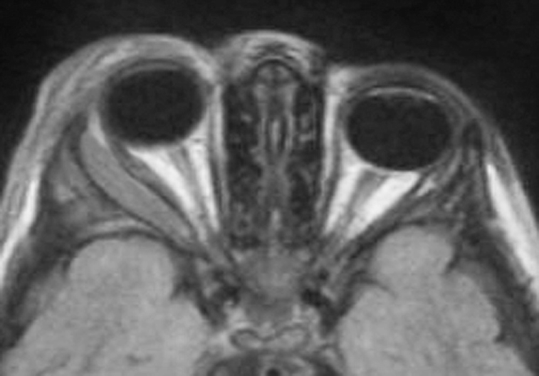
Orbital Tumors Springerlink

Malassezia Pachydermatis An Overview Sciencedirect Topics
Https Link Springer Com Content Pdf 10 1007 2f978 1 4614 7849 2 Pdf
Http Link Springer Com Content Pdf 10 1007 2f978 1 4939 1477 7 Pdf
Https Jamanetwork Com Journals Jamadermatology Articlepdf 530375 Archderm 96 3 004 Pdf
Pathology Outlines Epidermal Epidermoid Type
Https Digital Library Adelaide Edu Au Dspace Bitstream 2440 57424 2 02whole Pdf
The Role Of Vitamin D In Melanogenesis With An Emphasis On Vitiligo Topic Of Research Paper In Clinical Medicine Download Scholarly Article Pdf And Read For Free On Cyberleninka Open Science

Adnexal Tumors Chapter 4 Pearls And Pitfalls In Neoplastic Dermatopathology